-45%   PROMOTION

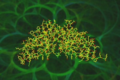
Papiers peints: Crystal structure of active human granzyme m. ribbons diagram

Papiers peints Crystal structure of active human granzyme M. Ribbons diagram in secondary structure coloring. Rendering based on protein data bank. Scientific background. 3d illustration
Papiers peints:
Crystal structure of active human granzyme M. Ribbons diagram in secondary structure coloring. Rendering based on protein data bank. Scientific background. 3d illustration.
Auteur: ©
Numéro de l'image:
#263711790
Autres sujets:
fond, coloré, bien-être, scientifique, recherche, la science, examens médicaux, en bonne santé, santé
Visualisation du produit
Similaire
Space-filling molecular model of human cathepsin G. Rendering based on protein data bank. Scientific background. 3d illustration
Space-filling molecular model of interleukin-38, a member of the interleukin-1 family and the interleukin-36 subfamily. Rendering based on protein data. 3d illustration
Crystal structure of cathepsin B in complex with nitroxoline. Ribbons diagram based on protein data bank entry 3ai8. Rainbow coloring from N to C. 3d illustration
Space-filling molecular model of human cathepsin G. Rendering based on protein data bank. Scientific background. 3d illustration
Molecular model of a newly discovered cytokine interleukin-38, a member of the interleukin-1 family and the interleukin-36 subfamily. Scientific background. 3d illustration
Crystal structure and molecular model of leptin, human peptide hormone, hunger or obesity hormone. Rendering based on protein data bank entry 1ax8. 3d illustration
Space-filling molecular model of canakinumab, a monoclonal antibody neutralizing interleukin-1beta. Rendering with differently colored protein chains based on protein data bank. 3d illustration
Molecular model of human pepsin 3b, one of the enzymes that digest food proteins into peptides. Rendering based on protein data bank entry 3utl Rainbow coloring from N to C. 3d illustration
Structural chemical formula and molecular model of beta-carotene isolated on white background. It is converted into vitamin A, an essential vitamin. 3d illustration
Crystal structure and space-filling molecular model of SARS-CoV-2 spike receptor-binding domain bound with ACE2. 3d illustration
Space-filling molecular model of human cathepsin B in complex with nitroxoline. Rendering based on protein data bank. Scientific background. 3d illustration
Molecular model of beta-sitosterol. Atoms are represented as spheres with conventional color coding: carbon (grey), oxygen (red), hydrogen (white). 3d illustration
Molecular model of recombinant human interleukin-22, a cytokine that acts on non-hematopoietic cells. Rendering with differently colored protein chains based on protein data bank. 3d illustration
Crystal structure and space-filling molecular model of interleukin-38, a member of the interleukin-1 family. Rainbow coloring from N to C. 3d illustration
Crystal structure of the tetanus neurotoxin produced by clostridium tetani bacteria. Scientific background. 3d illustration
Space-filling molecular model of human cathepsin B in complex with nitroxoline. Rendering with differently colored protein chains based on protein data bank. 3d illustration
Crystal structure of human cathepsin D in complex with macrocyclic inhibitor 14. Ribbons diagram with multi-colored protein chains based on protein data bank. 3d illustration
Crystal structure and molecular model of the human granzyme B isolated on white background. Rendering based on protein data bank. 3d illustration
Glycogen molecule, a polysaccharide that serves as a form of energy storage of animals, fungi and bacteria. Scientific background. 3d illustration
Human cathepsin G. Atoms are shown as spheres with conventional color coding: carbon (grey), oxygen (red), hydrogen (white), nitrogen (blue), sulfur (yellow), phosphorus (orange). 3d illustration
Crystal structure and molecular model of human cathepsin B in complex with nitroxoline. Rendering based on protein data bank. Scientific background. 3d illustration
Molecular model of a newly discovered cytokine interleukin-38, a member of the interleukin-1 family and the interleukin-36 subfamily. Rendering based on protein data bank. 3d illustration
Crystal structure of the pro-inflammatory cytokine interleukin 36 gamma. Ribbons diagram in secondary structure coloring based on protein data bank. Scientific background. 3d illustration
Space-filling molecular model of the insulin receptor ectodomain in complex with one insulin molecule isolated on white background. Medical background. Scientific background. 3d illustration
Space-filling molecular model of human interleukin-15 quaternary complex isolated on white background. Rendering with differently colored protein chains based on protein data bank. 3d
Crystal structure and space-filling molecular model of human interleukin 1 beta. Rendering based on protein data bank. Rainbow coloring from N to C. Scientific background. 3d illustration
Space-filling molecular model of anticalin-colchicine complex. Anticalins are artificial proteins that are able to bind to antigens, either to proteins or small molecules. 3d illustration
Crystal structure and molecular models of human interleukin-6, pro-inflammatory cytokine and anti-inflammatory myokine. Rendering based on protein data bank. 3d illustration
Crystal structure of the pro-inflammatory cytokine interleukin 36 gamma. Ribbons diagram in secondary structure coloring based on protein data bank. Scientific background. 3d illustration
Space-filling molecular model of human cathepsin D in complex with macrocyclic inhibitor 14. Rendering with differently colored protein chains based on protein data. 3d illustration
Crystal structure of an anticalin-colchicine complex. Ribbons diagram based on protein data bank. Rainbow coloring from N to C. Scientific background. 3d illustration
Crystal structure and molecular model of human intelectin-1, also known as omentin. Rendering with differently colored protein chains based on protein data bank. 3d illustration
Molecular model of interleukin 36 gamma, previously known as interleukin-1 family number 9, a protein that in humans encoded by the IL36G gene. 3d illustration
Crystal structure of anticalin-colchicine complex. Ribbons diagram in secondary structure coloring based on protein data bank entry 5nkn. Scientific background. 3d illustration
Crystal structure and molecular model of human cathepsin D in complex with macrocyclic inhibitor 14 isolated on white background. 3d illustration
Molecular model of the pro-inflammatory cytokine interleukin 36 gamma. Rendering based on protein data. Rainbow coloring from N to C. Scientific background. 3d illustration
Molecular model of human cathepsin B in complex with nitroxoline. Cathepsin B is a protein that in humans encoded by the CTSB gene. 3d illustration
Crystal structure of human cathepsin D in complex with macrocyclic inhibitor 14. Ribbons diagram in secondary structure coloring based on protein data bank. 3d illustration
Space-filling molecular model of the human granzyme B, a protein released by pathogen-infected or transformed target cells during cellular immune reactions. 3d illustration
Crystal structure and molecular model of interleukin-36 gamma, previously known as interleukin-1 family number 9. Scientific background . 3d illustration
Crystal structure of alpha-galactosidase enzyme that causes Fabry disease. Ribbons diagram in rainbow colors. Scientific background. 3d illustration
Crystal structure and space-filling molecular model of human pancreatic alpha-amylase. Rendering based on protein data bank. Rainbow coloring from N to C. 3d illustration
Molecular model of interleukin-36 alpha isolated on white background. Rendering based on protein data bank entry 6hpi. Rainbow coloring from N to C. 3d illustration
Structural chemical formula and space-filling molecular model of mirtazapine, an antidepressant medicine used to treat depression, compulsive and anxiety disorders. 3d illustration
Space-filling molecular model of human interleukin-18 (IL-18) receptor complex. Rendering with differently colored protein chains based on protein data bank entry 4r6u. 3d illustration
Molecular model of recombinant human interleukin-22. Rendering based on protein data bank entry 1m4r. Rainbow coloring from N to C. Scientific background. 3d illustration
Molecular structure of the N-terminal domain of carcinoembryonic antigen (CEA). Rendering based on protein data bank entry 2qsq. Scientific background. 3d illustration
Crystal structure of human interleukin-8 in solution. Ribbons diagram in secondary structure coloring. Scientific background. 3d illustration
Human interleukin-7 with unglycosylated interleukin-7 receptor alpha ectodomain. Scientific background. 3d illustration
Crystal structure of recombinant human interleukin-22. Ribbons diagram in secondary structure coloring. Rendering based on protein data bank entry 1m4r. 3d illustration
Crystal structure of human interleukin-3 in complex with receptor. Ribbons diagram with differently colored protein chains based on protein data bank entry 5uv8. 3d illustration
Molecular structure of human chorionic gonadotropin, a hormone produced by placenta during pregnancy. Rendering with multi-colored protein chains based on protein data bank. 3d illustration
Space-filling molecular model of the human Complement C3b in complex with Smallpox Inhibitor of Complement. Rendering based on protein data bank. Scientific background. 3d illustration
Molecular model of acetone or propanone, a colorless volatile and flammable liquid with a characteristic pungent odour, used in manufacture of plastics and other industrial products. 3d illustration
Molecular structure of adapalene, a third-generation topical retinoid used to treat mild to moderate acne, hair keratosis, and other skin conditions. Scientific background. 3d illustration
Space-filling molecular model of naphthalene. Atoms are represented as spheres with conventional color coding: carbon (grey), hydrogen (white). 3d illustration
Space-filling and ball-and-stick molecular models of human interleukin-6 or IL-6, pro-inflammatory cytokine and anti-inflammatory myokine. 3d illustration
Molecular model of human anaplastic lymphoma kinase (ALK) in complex with crizotinib. ALK is a receptor tyrosine kinase and enzyme that in humans is encoded by the ALK gene. 3d illustration
Crystal structure of human methionine aminopeptidase-2, an enzyme that in humans is encoded by the METAP2 gene. Ribbons diagram in rainbow colors. 3d illustration
Molecular model of thymine one of the four main bases found in DNA and RNA, along with adenine, cytosine and guanine. Scientific background. 3d illustration
Space-filling molecular model of interleukin-36 gamma, previously known as interleukin-1 family number 9, a protein that in humans encoded by the IL36G gene. 3d illustration
Crystal structure of cathepsin K with a covalently-linked inhibitor. Ribbons diagram based on protein data bank. Rainbow coloring from N to C. Scientific background. 3d illustration
Molecular model of human cathepsin D in complex with macrocyclic inhibitor 14. Rendering based on protein data bank entry 6qbg. 3d illustration
Crystal structure and molecular model of anticalin-colchicine complex. Rendering based on protein data bank. Scientific background. 3d illustration
Molecular model of anticalin-colchicine complex. Anticalins are artificial proteins that are able to bind to antigens, either to proteins or small molecules. Scientific background. 3d illustration
Space-filling molecular model of human cathepsin D in complex with macrocyclic inhibitor 14. Rendering based on protein data bank. Scientific background. 3d illustration
Crystal structure of human (IL-21) in complex with interleukin-21 receptor (IL21R). Ribbons diagram with multi-colored protein chains based on protein data bank. 3d illustration
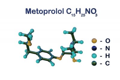
Structural chemical formula and molecular model of metoprolol, a beta-blocker used in treatment of angina pectoris, heart failure, and high blood pressure. Scientific background. 3d illustration
Molecular model of guanine one of the four main bases found in DNA and RNA, along with adenine, cytosine and thymine. Scientific background. 3d illustration
Molecular model of human prolactin. Atoms are represented as spheres with conventional color coding: carbon (grey), hydrogen (white), oxygen (red), nitrogen (blue), sulfur (yellow). 3d illustration
Structural chemical formula and space-filling molecular model of ergosterol or provitamin D2, a sterol found in cell membranes of fungi and protozoa. 3d illustration
Structural chemical formula and space-filling molecular model of arachidonic acid, a polyunsaturated fatty acid that consumed in our regular diets. Scientific background. 3d illustration
Structural chemical formula and space-filling molecular model of rutin, a bioflavonoid found in certain vegetables and fruits that has powerful antioxidant properties. 3d illustration
Structural chemical formula and molecular model of tryptamine, a monoamine alkaloid, structurally similar to the amino acid tryptophan. 3d illustration
Structural chemical formula and space-filling molecular model of luteolin, a yellow crystalline flavonoid widely present in fruits, vegetables and medicinal herbs. 3d illustration
Structural chemical formula and molecular model of l-carnosine, a new active ingredients for food supplement, which is widely used in food and can be used also in cosmetics. 3d illustration
Molecular model of human salivary amylase, a glucose-polymer cleavage enzyme that produced by the salivary glands. Scientific background. 3d illustration
Molecular structure of astaxanthin, keto-carotenoid that responsible of pink-red color of salmon and shrimps, antioxidant food supplement, e 161j. 3d illustration
Structural chemical formula and molecular model of limonene, the major component in the oil citrus fruit peels. 3d illustration
Molecular model of amphetamine. Atoms are represented as spheres with conventional color coding: carbon (grey), nitrogen (blue), hydrogen (white). 3d illustration
voir plusgarder
Matériaux

Papier peint vinyle

Le papier peint vinyle est imprimé sur un support résistant à la une surface lisse et mate.

La technologie HP Latex utilisée vous fera profiter de couleurs intenses au fil du temps.

  • ✓ Résistant à la déformation et à l'étirement
  • ✓ Perméable à la vapeur d'eau
  • ✓ Facile à poser
  • ✓ Encre HP Latex écologique

Largeur maximale des lés de papier peint : 95-105 cm. Pour les dimensions supérieures à la largeur du matériau, les impressions seront réalisées sur plusieurs lés égaux
Utilisation : murs lisses et plats
Collage: bord à bord. La colle ne s’applique que sur la surface de vos murs ce qui réduit le risque d'endommagement de votre papier peint et permet également de petites corrections afin d’ajuster la pose
Nettoyage : chiffon sec
Finition : semi-mate


Papier peint vinyle lavable

Le papier peint vinyle lavable est imprimé sur un support résistant à la une surface lisse et mate. Il est également stratifié ce qui le protège de l’humidité et autres facteurs extérieurs (éraflure, égratignure, exposition au soleil).

La technologie HP Latex utilisée vous fera profiter de couleurs intenses au fil du temps.

  • ✓ Stratifié
  • ✓ Résistant à l’humidité, aux éraflures, égratignures et à l’exposition au soleil
  • ✓ Résistant à la déformation et à l'étirement
  • ✓ Perméable à la vapeur d'eau
  • ✓ Facile à poser
  • ✓ Encre HP Latex écologique

Largeur maximale des lés de papier peint: 95-105 cm. Pour les dimensions supérieures à la largeur du matériau, les impressions seront réalisées sur plusieurs lés égaux
Utilisation : murs lisses et plats. Pièces avec une forte présence d’humidité (cuisine, salle de bains) ou celles dont les surfaces seraient susceptibles d’être éraflées (couloir, chambre d’enfant, hôtel, restaurant, salle de conférence, bâtiment public, école, maternelle, crèche)
Collage: bord à bord. La colle ne s’applique que sur la surface de vos murs ce qui réduit le risque d'endommagement de votre papier peint et permet également de petites corrections afin d’ajuster la pose
Nettoyage: chiffon humide
Finition: semi-mate

Papier peint vinyle expansé

Le papier peint vinyle est imprimé sur un support résistant à la structure « sable fin » ce qui intensifie le rendu de profondeur de l’impression. La technologie HP Latex utilisée vous fera profiter de couleurs intenses au fil du temps.

  • ✓ Structure sable fin
  • ✓ Résistant à la déformation et à l'étirement
  • ✓ Perméable à la vapeur d'eau
  • ✓ Facile à poser
  • ✓ Encre HP Latex écologique

Largeur maximale des lés de papier peint: 100cm. Pour les dimensions supérieures à la largeur du matériau, les impressions seront réalisées sur plusieurs lés égaux
Utilisation : murs lisses et plats. Le papier peint photo conviendra aussi bien dans des intérieurs comme la maison mais aussi dans un restaurant, une l’école ou encore au travail dans les bureaux
Collage: bord à bord. La colle ne s’applique que sur la surface de vos murs ce qui réduit le risque d'endommagement de votre papier peint et permet également de petites corrections afin d’ajuster la pose
Nettoyage: chiffon sec
Finition: semi-mate, structure sable fin


Papier peint vinyle expansé lavable

Le papier peint vinyle est imprimé sur un support résistant à la structure «sable fin» ce qui intensifie le rendu de profondeur de l’impression. La technologie HP Latex utilisée vous fera profiter de couleurs intenses au fil du temps.

  • ✓ Stratifié
  • ✓ Structure sable fin
  • ✓ Résistant à l’humidité, aux éraflures, égratignures et à l’exposition au soleil
  • ✓ Résistant à la déformation et à l'étirement
  • ✓ Perméable à la vapeur d'eau
  • ✓ Facile à poser
  • ✓ Encre HP Latex écologique

Largeur maximale des lés de papier peint: 100cm. Pour les dimensions supérieures à la largeur du matériau, les impressions seront réalisées sur plusieurs lés égaux
Utilisation : murs lisses et plats. Pièces avec une forte présence d’humidité (cuisine, salle de bains) ou celles dont les surfaces seraient susceptibles d’être éraflées (couloir, chambre d’enfant, hôtel, restaurant, salle de conférence, bâtiment public, école, maternelle, crèche)
Collage: bord à bord. La colle ne s’applique que sur la surface de vos murs ce qui réduit le risque d'endommagement de votre papier peint et permet également de petites corrections afin d’ajuster la pose
Nettoyage: chiffon humide
Finition: semi-mate, structure sable fin


Papier peint autocollant

Ce papier peint conviendra à différents types de pièces et de surfaces. Les papiers peints autocollants pourront décorer vos murs, vos armoires, vos vitres, vos meubles et autres surfaces lisses. La technologie HP Latex utilisée vous fera profiter de couleurs intenses au fil du temps.

  • ✓ Pas besoin de colle
  • ✓ Très facile à poser
  • ✓ Résistant à la déformation et à l'étirement
  • ✓ Perméable à la vapeur d'eau
  • ✓ Encre HP Latex écologique

Largeur maximale des lés de papier peint: 133cm. Pour les dimensions supérieures à la largeur du matériau, les impressions seront réalisées sur plusieurs lés égaux
Utilisation : surfaces lisses, utilisation universelle
Collage: bord à bord
Nettoyage: chiffon humide
Finition: semi-mate


voir plusgarder
Voir aussi
La planète Terre de l'espace
Texture abstrait abstraite colorée
Terre de l'espace
La planète Terre de l'espace
Terre de l'espace
La planète Terre de l'espace
Terre de l'espace
La planète Terre de l'espace
Texture abstrait abstraite colorée
La planète Terre de l'espace
Terre de l'espace
Terre de l'espace
Virus de l'hépatite C sur fond bleu, illustration 3D
Texture abstrait abstraite colorée
Virus de l'hépatite C sur fond rouge, illustration 3D
Virus de l'hépatite B sur fond coloré, illustration 3D
Virus de l'hépatite C sur fond coloré, illustration 3D
Rotavirus, un virus ARN, illustration 3D
Fond transparent avec eau de mer transparente
Rotavirus, un virus ARN, illustration 3D
Hiv, aides virus et érythrocytes isolé sur fond noir, illustration 3D
virus dans le sang vert, illustration 3D
Hiv, aide les virus dans le sang, illustration 3D
Virus bleu dans le sang, illustration 3D
Virus de l'hépatite C dans le sang, illustration 3D
Virus de l'hépatite B dans le sang, illustration 3D
Virus de l'hépatite C dans le sang, illustration 3D
Virus dans le sang, illustration 3D
Virus de l'hépatite C dans le sang, illustration 3D
Terre de l'espace
Virus et bactéries dans le sang, illustration 3D
Virus bleu sur fond coloré, illustration 3D
Virus et bactéries sur fond coloré, illustration 3D
Virus et bactéries dans le sang, illustration 3D
Mycobactéries isolées sur fond noir, illustration 3D
Bactérie tuberculeuse isolée sur fond noir, illustration 3D
Virus de l'herpès isolé sur fond noir, illustration 3D
Virus de l'herpès dans le sang, illustration 3D
Virus et bactéries sur fond bleu, illustration 3D
Virus du sang, illustration 3D
Virus sur fond bleu, illustration 3D
Virus et bactéries sur fond bleu, illustration 3D
Virus et bactéries sur fond coloré, illustration 3D
Virus pathogènes humains, illustration 3D
Contexte scientifique. La loi intégrale de conservation de masse, illustration 3D
Virus et bactéries pathogènes humaines, illustration 3D
Contexte scientifique. La loi intégrale de conservation de masse, illustration 3D
Virus pathogènes humains et bactéries, illustration 3d
Virus sur fond bleu, illustration 3D
Virus sur fond coloré, illustration 3D
Virus sur fond rouge, illustration 3D
Virus et bactéries isolés sur fond noir, illustration 3D
Virus et bactéries dans le sang, illustration 3D
Bactéries sur fond bleu, illustration 3D
Bactéries sur fond rouge, illustration 3D
Bactéries sur fond coloré, illustration 3D
Bactéries sur fond vert, illustration 3D
voir plusgarder

Notre boutique en ligne utilise des cookies et des technologies connexes. Grâce à eux, nous assurons une qualité de service optimale et garantissons le bon fonctionnement du site web. Une fois votre consentement obtenu, les cookies seront également utilisés pour des mesures supplémentaires et l'analyse de l'utilisation du site. Cela nous permettra d'adapter le site à vos intérêts et de vous présenter un contenu et une publicité personnalisés.
Vos données sont gérées par Fairbox Sp. z o.o. et, dans certaines situations, par nos partenaires avec lesquels nous coopérons. Pour des informations détaillées sur l'utilisation des cookies et le traitement de vos données personnelles, veuillez consulter notre Politique en matière de confidentialité et de cookies.
Paramètres des cookies
Vous trouverez ci-dessous les descriptions des différentes catégories de cookies et la possibilité de les personnaliser selon vos préférences:
Les cookies indispensables
Nous les utilisons pour assurer le bon fonctionnement du site web. Ils vous permettent de naviguer dans notre boutique sans interruption et d'effectuer vos achats en toute sécurité.
Les cookies fonctionnels
Ils sont utilisés pour mémoriser les choix que vous faites sur le site, ce qui permet d'adapter le site à vos préférences personnelles et d'améliorer votre expérience en tant qu'utilisateur.
Les cookies analytiques
Les cookies analytiques mesurent le nombre de visites et collectent des informations sur les sources de trafic du site. Ils nous permettent de comprendre comment les utilisateurs se servent du site, d'améliorer la qualité du service et d'adapter le contenu de la boutique pour mieux répondre aux besoins de nos clients.
Les cookies marketing
Les cookies marketing nous permettent, ainsi qu'à nos partenaires, d'adapter le contenu publicitaire à vos centres d'intérêt. Ils sont également utilisés pour afficher des publicités sur d'autres sites web.